CoIP, ChIP, CUT&RUN, CUT&Tag Expert! My Basket (...)
Worldwide delivery!
Having the Best Alpaca Nanobody Products!
Home | About us | Contact us | How to order |
    My Base Info
    Shopping Basket
    My Orders
    My Shipping Info
    Modify Password
    Log Out

    Anti-HA tag Mouse mAb

    Catalog number :AT0024
    loading...
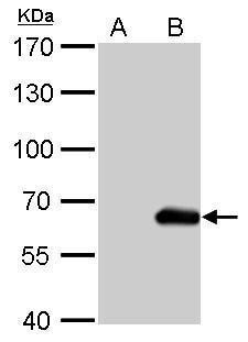
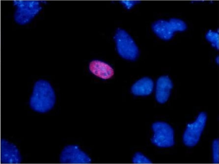
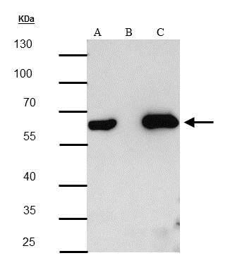
    The HA tag is derived from an epitope of the influenza hemagglutinin protein which has been used extensively as a general epitope tag in expression vectors.
    Human influenza hemagglutinin (HA) is a surface glycoprotein required for the infectivity of the human virus. The HA tag is derived from the HA molecule corresponding to amino acids 98-106 has been extensively used as a general epitope tag in expression vectors. Many recombinant proteins have been engineered to express the HA tag, which does not appear to interfere with the bioactivity or the biodistribution of the recombinant protein. This tag facilitates the detection, isolation, and purification of the proteins.
    Overview
    Description
    Mouse monoclonal antibody to HA tag - Epitope Tag Antibody
    Reactivity
    All Species Expected
    Tested applications
    WB    : 1/2000-1/10000.
    IHC    : 1/200-1/5000.
    ICC/IF    : 1/200-1/2000.
    IP/CoIP    : 1/100-1/500.
    Not yet tested in other applications. Optimal dilutions/concentrations should be determined by the end user.
    Product Picture
    Anti-HA tag Mouse mAb
    Specificity
    The antibody recognizes exogenously expressed proteins containing the HA epitope tag.
    Properties
    Immunogen
    YPYDVPDYA (influenza hemagglutinin-HA-epitope) conjugated to KLH.
    Clonality
    Monoclonal, clone number: 6A1
    Isotype
    Mouse IgG2a
    Form
    Liquid, 100 μg at 1mg/mL
    Mouse monoclonal immunoglobulin in phosphate buffered saline (PBS), pH 7.3 (+/- 0.1), 50% glycerol with 0.1mg/mL BSA (IgG, protease free) as a stabilizer and 0.02% sodium azide as preservative.
    Storage instruction
    Store at +4°C short term (1-2 weeks). Aliquot and store at -20°C, Avoid freeze / thaw cycle.
    Database links
    Sequence: YPYDVPDYA (influenza hemagglutinin-HA-epitope).
    Host
    Mouse
    Applications
    WB Image

    Western blot shows lysates of HEK293 human embryonic kidney cell line transfected with N-terminal HA-tagged LGI-2, Internal Sequence HA-tagged CIGALT, and C-terminal HA-tagged FBLN. PVDF membrane was probed with 1:5000 of Anti-HA Antibody (Catalog # AT0024, Engidody) followed by HRP-conjugated Anti-Mouse IgG Secondary Antibody (Catalog # AT0098, Engidody). Specific bands were detected for HA-tagged proteins at approximately 60, 37, and 150 kDa (as indicated).
    ICC/IF Image

    Immunocytochemistry/Immunofluorescence: HA Epitope Tag Antibody - Analysis of HA tag transfected HeLa cells were fixed in 4% paraformaldehyde at RT for 15 min.
    Red: HA-recombinant protein stained by HA tag antibody diluted at 1:1000.
    Blue: Hoechst 33342 staining.
    IP/CoIP Image

    Immunoprecipitation and Western Blot: Anti-HA Epitope Tag Antibody - Analysis of 293T whole cell lysate/extract
    A: 20 ug whole cell lysate/extract of HA tagged protein expressing 293T cells
    B: Control with 1:5000 of mouse normal IgG
    C: Immunoprecipitation of HA tagged protein by 1:200 of HA tag antibody, the immunoprecipitated HA tagged protein was detected by HA tag antibody diluted at 1:5000.
    Protocols
    Western Blotting Procedure
     
    1. Lyse approximately 107 cells in 0.5 mL of ice-cold Cell Lysis Buffer (formulation provided below). This buffer, a modified RIPA buffer, is suitable for recovery of most proteins, including membrane receptors, cytoskeletal-associated proteins, and soluble proteins. This cell lysis buffer formulation is available as a separate product which requires supplementation with protease inhibitors immediately prior to use (Cat. # FNN0011, thermofisher). Other cell lysis buffer formulations, such as Laemmli sample buffer and Triton-X 100 buffer, are also compatible with this procedure. Additional optimization of the cell stimulation protocol and cell lysis procedure may be required for each specific application.
     
    2. Remove the cellular debris by centrifuging the lysates at 14,000 x g for 10 minutes. Alternatively, lysates may be ultra-centrifuged at 100,000 x g for 30 minutes for greater clarification.
     
    3. Carefully decant the clarified cell lysates into clean tubes and determine the protein concentration using a suitable method, such as the Bradford assay. Polypropylene tubes are recommended for storing cell lysates.
     
    4. React an aliquot of the lysate with an equal volume of 2x Laemmli Sample Buffer (125 mM Tris, pH 6.8, 10% glycerol, 10% SDS, 0.006% bromophenol blue, and 130 mM dithiothreitol [DTT]) and boil the mixture for 90 seconds at 100°C.
     
    5. Load 10-30 µg of the cell lysate into the wells of an appropriate single percentage or gradient mini-gel and resolve the proteins by SDS-PAGE.
     
    6. In preparation for the Western transfer, cut a piece of PVDF slightly larger than the gel. Soak the membrane in methanol for 1 minute, then rinse with ddH2O for 5 minutes. Alternatively, nitrocellulose may be used.
     
    7. Soak the membrane, 2 pieces of Whatman paper, and Western apparatus sponges in transfer buffer (formulation provided below) for 2 minutes.
     
    8. Assemble the gel and membrane into the sandwich apparatus.
     
    9. Transfer the proteins at 140 mA for 6°C 0-90 minutes at room temperature.
     
    10. Following the transfer, rinse the membrane with Tris buffered saline for 2 minutes.
     
    11. Block the membrane with blocking buffer (formulation provided below) overnight at 4°C or for one hour at room temperature.
     
    12. Incubate the blocked blot with primary antibody at a 1:1000 starting dilution in Tris buffered saline supplemented with 1% Ig-free BSA and 0.1% Tween-20 overnight at 4°C or for two hours at room temperature.
     
    13. Wash the blot with several changes of Tris buffered saline supplemented with 0.1% Tween 20.
     
    14. Detect the primary antibody band using an appropriate secondary antibody, such as goat anti-rabbit IgG conjugated HRP (Cat. # AT0097-100ul, Engibody) or goat anti-mouse IgG conjugated HRP (Cat. # AT0098-100ul, Engibody) in conjunction with your chemiluminescence reagents and instrumentation.
    Troubleshooting Guide
    Western Blot Troubleshooting
    1. No signal
    2. High background
    3. Non specific bands
    4. Incorrect band size
    5. White bands on black/grey blot
    6. ‘Smear’
     
    1. Troubleshooting:  NO SIGNAL
    Sample preparation
    • Untested species? Positive control. Check alignment.
    • Not enough antigen in the sample or loaded onto the gel.
    Transfer to the membrane
    • Poor transfer to membrane.
    • Excessive washing.
    Blocking
    • Too much blocking. Optimize blocking agent, concentration and time
    • Cross-reaction blocking agent / antibody.
    Antibody incubation and detection
    • Not enough antibody.
    • Incorrect secondary antibody.
    • Detection kit is old - substrate inactive.
     
    2. Troubleshooting:  HIGH BACKGROUND
    Transfer to the membrane
    • Choice of membrane.
    Blocking
    • Need to block for longer.
    • Optimize blocking agent, concentration.
    • Cross-reaction between blocking agent and primary or secondary.
    • Phospho-specific proteins: use Casein to block.
    Antibody incubation and detection
    • Primary antibody concentration too high.
    • Incubation temperature too high.
     
    3. Troubleshooting:  NON SPECIFIC BANDS
    Sample preparation
    • Cell lines can accumulate differences in their protein expression profiles.
    • Protein has multiple modified forms, altering mobility.
    • Target protein is in fragments – degradation by proteases or cleaved.
    • Splice variants / isoforms or possibly proteins from the same family.
    • Multimers (dimers) of protein (reduce and denature).
    • Bands may be non-specific. Run a no primary control
     Blocking
    • Insufficient blocking. Optimizing concentration, time.
    Antibody incubation and detection
    • Primary or secondary antibody concentration too high.
    • Antibody has not been purified.
     
    4. Troubleshooting:  INCORRECT BAND SIZE
    Sample preparation
    • Ensure sample is reduced and denatured unless stated otherwise on the datasheet.
    • Check for isoforms.
    • Is the sample a recombinant fragment? These will run at a different size.
     
    5. Troubleshooting:  WHITE BANDS/ BLACK BLOT
    • Too much primary and/or too much secondary antibody.
     
    6. Troubleshooting: ‘Smear’
    • Overloading, Protein degradation.

    Related Products

    Catalog numberAT0057
    Catalog numberAT0023
    Catalog numberAT0025
    Catalog numberAT0026
    Catalog numberAT0027
    Catalog numberAT0028
    Catalog numberAT0029
    Catalog numberAT0030

    Reviews

    loading...
    Content *:
    Customer Review :
    Verification Code * :
    refresh image

    Shipping and Paying Info
    ......
    Order Information
    You can place an order online step by step as blow.

    Step 1. Register for a new account and Log in, Find out your product by searching our website.
    Step 2. Add the Product into your shopping basket, select your settlement currency.
    Step 3. Checkout by Paypal, Pay by credit card or bank transfer for your order.
    Step 4. Ship the goods for you, providing you package tracking numbers. 

    Any questions, please contact us via email: sale@engibodybio.com
    Payments by
    All our products are For Research Use Only. Not for diagnostic or therapeutic usages.