The H-H-H-H-H-H motif is used as a tag on many recombinant proteins to facilitate purification. The antibody recognizes the His-tag fused to the amino- or carboxy- termini of targeted proteins in transfected or transformed cells.
As is the case with other protein tag systems, this polyhistidine tag can often be cleaved at sites recognized by proteases such as thrombin and enterokinases to isolate the protein of interest.
Anti-His tag Mouse mAb
Catalog number :AT0025
- Overview
- Description
- Mouse monoclonal antibody to 6X His tag - Epitope Tag Antibody
- Reactivity
- All Species Expected
- Tested applications
- WB : 1/1000-1/5000.
IHC : 1/200-1/3000.
ICC/IF : 1/200-1/1000.
IP/CoIP : 1/100-1/500.
RIP : 1/100-1/200.
Not yet tested in other applications. Optimal dilutions/concentrations should be determined by the end user.
- Product Picture
- Specificity
- This antibody detects recombinant proteins containing the 6xHis epitope tag. The antibody recognizes the 6xHis-tag fused to either the amino or carboxy terminus of targeted proteins in transfected cells.
- Properties
- Immunogen
- Synthetic peptide: HHHHHH conjugated to KLH.
- Clonality
- Monoclonal, clone number: 2B6
- Isotype
- Mouse IgG1
- Form
- Liquid,100 μg at 1mg/mL
Mouse monoclonal immunoglobulin in phosphate buffered saline (PBS), pH 7.3 (+/- 0.1), 50% glycerol with 0.1mg/mL BSA (IgG, protease free) as a stabilizer and 0.02% sodium azide as preservative.
- Storage instruction
- Store at -20°C, Avoid freeze / thaw cycle.
- Database links
- Sequence: HHHHHH
- Host
- Mouse
- Applications
- WB Image
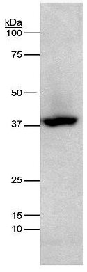
Lane 1: lysates prepared from HEK-293 cells transfected with his-tagged protein at 1:4000 dilutionRecommended secondary antibody: Goat anti mouse IgG-HRP (Engibody, Catalog number:AT0098)Recommended ECL: ECL Pico-Detect™ Western Chemiluminescent HRP Substrate (Engibody, Catalog number: IF6747)
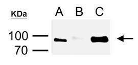
- WB Figure 1
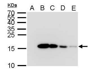
Western Blot:12 % SDS-PAGE 6x His antibody at 1:5000.A) 2 µg GST-recombinant proteinB) 2 µg His-recombinant proteinC) 1 µg His-recombinant proteinD) 0.5 µg His-recombinant proteinE) 0.1 µg His-recombinant protein.Recommended secondary antibody: Goat anti mouse IgG-HRP (Engibody, Catalog number:AT0098)Recommended ECL: ECL Pico-Detect™ Western Chemiluminescent HRP Substrate (Engibody, Catalog number: IF6747)
- ICC/IF Image
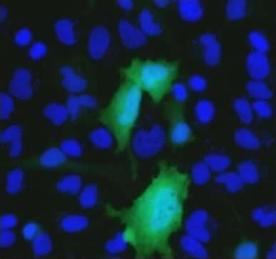
Immunocytochemistry/Immunofluorescence: - Analysis using Anti-6 x Histidine Epitope Tag Antibody and goat anti mouse IgG conjugated DyLight 488.Staining of HeLa cells transfected with a construct containing a 6x-His Epitope Tag. Formalin fixed cells were permeabilized with 0.1% Triton X-100 in TBS for 10 minutes at room temperature and blocked with 1% Blocker BSA for 15 minutes at room temperature. Cells were probed with Anti-6x-His Epitope Tag mouse monoclonal antibody at a dilution of 1:500 for at least 1 hour at room temperature, then using goat anti mouse IgG conjugated DyLight 488. Nuclei (blue) were stained with Hoechst 33342 dye.
- IP/CoIP Image
Immunoprecipitation and Western Blot: His Tag Antibody - Analysis of cell lysate/extract of His-fused gene transfected HeLa cellsA. 20µg lysate/extract of transfected HeLa cellsB. Control with 1:1000 of mouse normal IgGC. Immunoprecipitation of His-fused protein by 1:500 of 6X His tag Mouse mAb, the immunoprecipitated His-fused protein was detected by 6X His tag Rabbit pAb diluted at 1:3000.Recommended immunoprecipitation antibody for IP: Anti-His tag Mouse mAb (Engibody, Catalog number:AT0025)Recommended protein A/G beads: Protein A/G Agarose Beads (Engibody, Catalog number: IF0001), or Protein A/G Magnetic Beads (Engibody, Catalog number: IF0002)Recommended detection antibody for downstream WB: Anti-His tag Rabbit mAb (Engibody, Catalog number:AT0042), so that the heavy and light chains from immunoprecipitation antibody can't be seen in WB.Recommended secondary antibody for downstream WB: Goat anti rabbit IgG-HRP (Engibody, Catalog number:AT0097)Recommended ECL: ECL Pico-Detect™ Western Chemiluminescent HRP Substrate (Engibody, Catalog number: IF6747)
- Protocols
- Western Blotting Procedure
1. Lyse approximately 107 cells in 0.5 mL of ice-cold Cell Lysis Buffer (formulation provided below). This buffer, a modified RIPA buffer, is suitable for recovery of most proteins, including membrane receptors, cytoskeletal-associated proteins, and soluble proteins. This cell lysis buffer formulation is available as a separate product which requires supplementation with protease inhibitors immediately prior to use (Cat. # FNN0011, thermofisher). Other cell lysis buffer formulations, such as Laemmli sample buffer and Triton-X 100 buffer, are also compatible with this procedure. Additional optimization of the cell stimulation protocol and cell lysis procedure may be required for each specific application.
2. Remove the cellular debris by centrifuging the lysates at 14,000 x g for 10 minutes. Alternatively, lysates may be ultra-centrifuged at 100,000 x g for 30 minutes for greater clarification.
3. Carefully decant the clarified cell lysates into clean tubes and determine the protein concentration using a suitable method, such as the Bradford assay. Polypropylene tubes are recommended for storing cell lysates.
4. React an aliquot of the lysate with an equal volume of 2x Laemmli Sample Buffer (125 mM Tris, pH 6.8, 10% glycerol, 10% SDS, 0.006% bromophenol blue, and 130 mM dithiothreitol [DTT]) and boil the mixture for 90 seconds at 100°C.
5. Load 10-30 µg of the cell lysate into the wells of an appropriate single percentage or gradient mini-gel and resolve the proteins by SDS-PAGE.
6. In preparation for the Western transfer, cut a piece of PVDF slightly larger than the gel. Soak the membrane in methanol for 1 minute, then rinse with ddH2O for 5 minutes. Alternatively, nitrocellulose may be used.
7. Soak the membrane, 2 pieces of Whatman paper, and Western apparatus sponges in transfer buffer (formulation provided below) for 2 minutes.
8. Assemble the gel and membrane into the sandwich apparatus.
9. Transfer the proteins at 140 mA for 6°C 0-90 minutes at room temperature.
10. Following the transfer, rinse the membrane with Tris buffered saline for 2 minutes.
11. Block the membrane with blocking buffer (formulation provided below) overnight at 4°C or for one hour at room temperature.
12. Incubate the blocked blot with primary antibody at a 1:1000 starting dilution in Tris buffered saline supplemented with 1% Ig-free BSA and 0.1% Tween-20 overnight at 4°C or for two hours at room temperature.
13. Wash the blot with several changes of Tris buffered saline supplemented with 0.1% Tween 20.
14. Detect the primary antibody band using an appropriate secondary antibody, such as goat anti-rabbit IgG conjugated HRP (Cat. # AT0097-100ul, Engibody) or goat anti-mouse IgG conjugated HRP (Cat. # AT0098-100ul, Engibody) in conjunction with your chemiluminescence reagents and instrumentation.
- Troubleshooting Guide
- Western Blot Troubleshooting
1. No signal
2. High background
3. Non specific bands
4. Incorrect band size
5. White bands on black/grey blot
6. ‘Smear’
1. Troubleshooting: NO SIGNAL
Sample preparation
• Untested species? Positive control. Check alignment.
• Not enough antigen in the sample or loaded onto the gel.
Transfer to the membrane
• Poor transfer to membrane.
• Excessive washing.
Blocking
• Too much blocking. Optimize blocking agent, concentration and time
• Cross-reaction blocking agent / antibody.
Antibody incubation and detection
• Not enough antibody.
• Incorrect secondary antibody.
• Detection kit is old - substrate inactive.
2. Troubleshooting: HIGH BACKGROUND
Transfer to the membrane
• Choice of membrane.
Blocking
• Need to block for longer.
• Optimize blocking agent, concentration.
• Cross-reaction between blocking agent and primary or secondary.
• Phospho-specific proteins: use Casein to block.
Antibody incubation and detection
• Primary antibody concentration too high.
• Incubation temperature too high.
3. Troubleshooting: NON SPECIFIC BANDS
Sample preparation
• Cell lines can accumulate differences in their protein expression profiles.
• Protein has multiple modified forms, altering mobility.
• Target protein is in fragments – degradation by proteases or cleaved.
• Splice variants / isoforms or possibly proteins from the same family.
• Multimers (dimers) of protein (reduce and denature).
• Bands may be non-specific. Run a no primary control
Blocking
• Insufficient blocking. Optimizing concentration, time.
Antibody incubation and detection
• Primary or secondary antibody concentration too high.
• Antibody has not been purified.
4. Troubleshooting: INCORRECT BAND SIZE
Sample preparation
• Ensure sample is reduced and denatured unless stated otherwise on the datasheet.
• Check for isoforms.
• Is the sample a recombinant fragment? These will run at a different size.
5. Troubleshooting: WHITE BANDS/ BLACK BLOT
• Too much primary and/or too much secondary antibody.
6. Troubleshooting: ‘Smear’
• Overloading, Protein degradation.
Related Products
| Catalog number | AT0051 |
| Catalog number | AT0084 |
| Catalog number | AT0085 |
| Catalog number | AT0090 |
| Catalog number | AT0022 |
| Catalog number | AT0023 |
| Catalog number | AT0024 |
| Catalog number | AT0026 |
| Catalog number | AT0027 |
| Catalog number | AT0028 |
| Catalog number | AT0029 |
Reviews
loading...