Attention: Agarose Beads should be resuspended well before used in IP, Co-IP and protein purification.
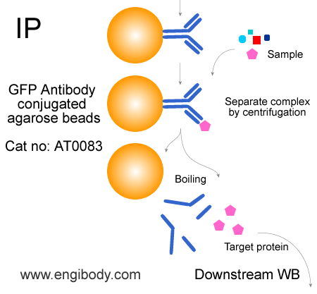
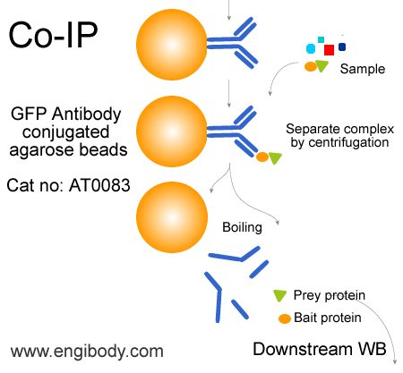
Protocol for Immunoprecipitation (IP/CoIP) of GFP-Fusion Proteins from mammalian cell lysate (For soluble cytoplasmic protein)
Solutions and Reagents
1X Cell Lysis Buffer: 50 mM Tris (pH 7.5), 150 mM NaCl, 1 mM EDTA, 1 mM EGTA, 1% Triton X-100. Recommend adding 1 mM PMSF before use.
1X Wash Buffer: TBS (50 mM Tris HCl, 150 mM NaCl, pH 7.5)
5X SDS Sample Loading Buffer
Procedure
Attention:
1. If your target protein is soluble nuclear protein in nucleus (Excluding nuclear membrane integrated protein and DNA-binding protein), please use NE-PER Nuclear and Cytoplasmic Extraction kit (Thermo, Cat no:78833) to extract nuclear protein. Or please use RIPA lysis buffer to lyse the cell, and add PIC, 1mM PMSF, 2.5mM Mgcl2 and 1mg/ml DNase I.
2. It is required to reserve 50 µl of protein extract for each sample as the INPUT control, which is directly used in the downstream WB experiment.
1. Harvest cells
1). For one immunoprecipitation reaction the use of ~106 - 107 mammalian cells (approx. one 10-cm dish) expressing a GFP-tagged protein of interest is recommended. To harvest adherent cells, aspirate growth medium, add 1 mL ice-cold PBS to cells and scrape cells from dish. Transfer cells to a pre-cooled tube, spin at 500 g for 3 min at +4°C and discard supernatant. Wash cell pellet twice with ice-cold PBS, gently resuspending the cells.
2. Lyse cells
1). Resuspend cell pellet in 500 µL ice-cold lysis buffer by pipetting.
Note: Add protease inhibitors and 1 mM PMSF into lysis buffer.
2). Place the tube on ice for 30 min with extensively pipetting every 10 min.
3). Centrifuge cell lysate at 14000x g for 10 min at +4°C. Transfer lysate(supernatant) to a pre-cooled tube. Discard pellet.
Note: At this point cell lysate may be put at -80°C for long-term storage.
3. Equilibrate beads
1). Resuspend the beads by inverting the product tube or gently pipetting up and down. Do not vortex the agarose beads,
2). Pipette 25 µL bead slurry into 500 µL ice-cold cell extract. Centrifuge at 2500x g for 2 min at +4°C. Discard supernatant and repeat wash twice.
4. Bind proteins
1). Add 25 µL equilibrated anti-GFP agarose beads slurry (step 3) into 500 µL cell extract (step 2). Save 30 µL of cell extract for input of immunoblot analysis before IP. Incubation at a rotator for 1-3 hours at 4°C or overnight 4°C.
2). Centrifuge at 2500x g for 2 min at +4°C. Discard supernatant.
5. Wash beads
1). Resuspend protein-antibody-beads complex in 1mL ice-cold wash buffer. Centrifuge at 2500x g for 5 min at +4°C. Discard supernatant and repeat wash at least three times.
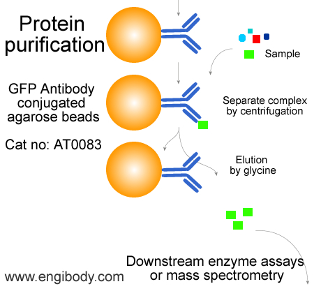
6. Elute proteins
Two elution methods are recommended according to protein characteristics or further usage:
Option A: Native Elution
Elution under acidic conditions with 0.2 M glycine (pH 2.5). This is a fast and efficient elution method. Neutralization of the eluted proteins with neutralizing buffer (1 M Tris, pH 10.4) may help preserve its activity. Neutralizing buffer needs to be placed in the collection tube in advance.
Attention: It is necessary to make a preliminary experiment in advance to determine how much neutralization buffer is needed to neutralize glycine (PH 2.5)
Option B: Denaturing Elution for SDS-PAGE
Elution with sample loading buffer under denaturing conditions for gel electrophoresis and immunoblotting.
A: Elution with 0.2 M Glycine (pH 2.5) - The procedure should be performed at room temperature.
Note:Do not leave the beads in this buffer more than 20 minutes.
1. Add 50 µL of 0.2 M Glycine (pH 2.5), to each sample and control beads complexes.
2. Incubate the samples and controls with constantly pipette up and down for 1-3 minutes at room temperature. (Note: Do not turn the tube upside down to prevent the complex from sticking to the tube wall)
3. Centrifugate the beads complex at 2500x g for 5 min at 4°C. Transfer the supernatants to fresh tubes containing about 5 µL of neutralizing buffer. Be careful not to transfer any beads.
4. Repeat steps 1 – 3 in order to improve elution efficiency, pooling eluates in same tube or collect eluate through different tubes.
6. For immediate use, store the eluates at 4 °C. Store at –20 °C for long term storage.
B: Elution with SDS-PAGE Sample Loading Buffer
1. Resuspend each sample with 30 µL 1X SDS sample loading buffer (6 µL 5X SDS sample loading buffer can be added into 24 µL cell lysis buffer). Vortex.
2. Boil the sample and control tubes for 10 minutes at 95 – 100°C to dissociate immunocomplex from anti-GFP agarose beads.
3. Anti-GFP agarose beads immunocomplex can be centrifugated at 2500x g for 2 min at 4°C. Transfer the supernatants to fresh tubes. The samples and controls are ready for loading on SDS-PAGE and immunoblotting using anti-GFP or specific antibodies against the fusion protein.