CoIP, ChIP, CUT&RUN, CUT&Tag Expert! My Basket (...)
Worldwide delivery!
Having the Best Alpaca Nanobody Products!
Home | About us | Contact us | How to order |
    My Base Info
    Shopping Basket
    My Orders
    My Shipping Info
    Modify Password
    Log Out

    Anti-Lamin A + C rabbit mAb - Nuclear Loading Control

    Catalog number :AT0797
    loading...
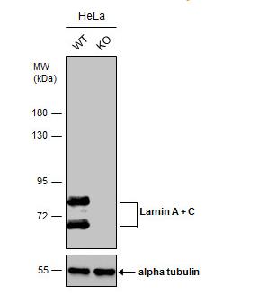
    LMNA gene conducts alternative splicing to produce Lamin A protein and Lamin C protein.
     
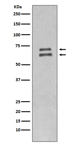
    Lamins are nuclear membrane structural components that are important in maintaining normal cell functions such as cell cycle control, DNA replication, and chromatin organization. Lamin A/C is cleaved by caspase-6 and serves as a marker for caspase-6 activation. During apoptosis, lamin A/C is specifically cleaved into a large (41-50 kDa) and a small (28 kDa) fragment. The cleavage of lamins results in nuclear dysregulation and cell death.
     
    Database Links
    UniProt ID: P02545 LMNA
    Entrez-Gene ID: 4000 LMNA
    Overview
    Reactivity
    Mouse, Rat, Human
    Tested applications
    WB : 1/1000-1/5000. 
    Predicted molecular weight: 74kDa (Lamin A)/63kDa (Lamin C), 28 kDa(The cleaved smaller fragments).
    IHC: 1/50~1/200
    ICC/IF: 1/50~1/200
    IP: 1/50
    FC: 1/50
    Optimal dilutions/concentrations should be determined by the end user.
     
    Specificity
    This antibody recognizes endogenous levels of 74kDa (Lamin A) and 63kDa (Lamin C). It also reacts with the smaller fragments of lamin A (28 kDa) and lamin C (28 kDa) produced by caspase 6 cleavage during apoptosis. This antibody does not cross-react with lamins B1 and B2.
    Properties
    Immunogen
    a recombinant fragment of human lamin A protein.
    Clonality
    Monoclonal, clone number: 3G1
    Isotype
    Rabbit IgG
    Form
    Liquid, 100 μl,1mg/ml, PBS (pH 7.2) and 40% Glycerol,0.02% Sodium Azide
    Storage instruction
    Store at -20°C, Avoid freeze / thaw cycle. Do not aliquot the antibody.
    Host
    Rabbit
    Applications
    WB Image

    Western Blotting Image
    Predicted band size: 74kDa (Lamin A) and 63kDa (Lamin C)
     
    Anti-Lamin B1 antibody at 1:2000
     
    Western Blot: Lamin A + C Antibody - Wild-type (WT) and Lamin A + C knockout (KO) HeLa cell extracts (30 ug) were separated by 7.5% SDS-PAGE, and the membrane was incubated with Lamin A + C antibody. The HRP-conjugated anti-rabbit IgG antibody was used to detect the primary antibody.
     
    Recommended ECL: ECL Pico-Detect™ Western Chemiluminescent HRP Substrate (Engibody, Catalog number: IF6747)

    WB Figure 2
    Anti- Lamin A/C antibody images: Western blotting.

    Western Blotting Image
    Predicted band size: 74kDa (Lamin A)/63kDa (Lamin C)
     
    Anti-Lamin A/C antibody at 1:2000, Cell lysates of Hela
     
    Recommended secondary antibody: Goat anti Rabbit IgG-HRP (Engibody, Catalog number:AT0097)
    Recommended ECL: ECL Pico-Detect™ Western Chemiluminescent HRP Substrate (Engibody, Catalog number: IF6747)

    Related Products

    Reviews

    loading...
    Content *:
    Customer Review :
    Verification Code * :
    refresh image

    Shipping and Paying Info
    ......
    Order Information
    You can place an order online step by step as blow.

    Step 1. Register for a new account and Log in, Find out your product by searching our website.
    Step 2. Add the Product into your shopping basket, select your settlement currency.
    Step 3. Checkout by Paypal, Pay by credit card or bank transfer for your order.
    Step 4. Ship the goods for you, providing you package tracking numbers. 

    Any questions, please contact us via email: sale@engibodybio.com
    Payments by
    All our products are For Research Use Only. Not for diagnostic or therapeutic usages.