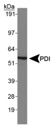
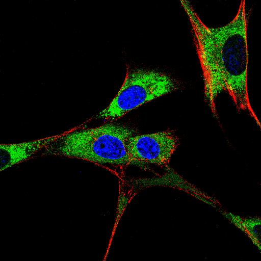
CoIP, ChIP, CUT&RUN, CUT&Tag Expert! My Basket (...)
Worldwide delivery!
Having the Best Alpaca Nanobody Products!
Home | About us | Contact us | How to order |
    My Base Info
    Shopping Basket
    My Orders
    My Shipping Info
    Modify Password
    Log Out

    Anti-PDI Rabbit mAb - ER Marker

    Catalog number :AT0793
    loading...
    Background: 
    During their synthesis, secretory proteins translocate into the endoplasmic reticulum (ER) where they are post-translationally modified and properly folded. To reach their native conformation, many secretory proteins require the formation of intra- or inter-molecular disulfide bonds. This process is called oxidative protein folding. Protein disulfide isomerase (PDI) catalyzes the formation and isomerization of these disulfide bonds. Studies on mechanisms of oxidative folding suggest that molecular oxygen oxidizes the ER-protein Ero1, which in turn oxidizes PDI through disulfide exchange. This event is then followed by PDI-catalyzed disulfide bond formation in folding proteins.
     
    UniProt ID: P07237
    Overview
    Reactivity
    Human, Mouse, Rat, Bovine, Pig, Monkey
    Tested applications
    Western Blotting: 1/1000-1/2000
    Immunofluorescence: 1/500
    Immunohistochemistry (Paraffin): 1/500
    Immunoprecipitation(IP) : 1/100 
    Optimal dilutions/concentrations should be determined by the end user. 
    Specificity
    This antibody detects endogenous levels of total PDI protein. 
    Properties
    Immunogen
    a synthetic peptide corresponding to the sequence of human PDI.
    Clonality
    Monoclonal, clone number: 6L7
    Isotype
    Rabbit IgG
    Form
    Liquid, 100 μl,1mg/ml, PBS (pH 7.2) and 40% Glycerol,0.02% Sodium Azide
    Storage instruction
    Store at +4°C short term (1-2 weeks). Aliquot and store at -20°C, Avoid freeze / thaw cycle.
    Host
    Rabbit
    Applications
    WB Image

    Western Blot: PDI Antibody - Normal human brain.
    ICC/IF Image

    Immunocytochemistry/Immunofluorescence: PDI Antibody - IF Confocal analysis of 3T3 cells using PDI antibody. An Alexa Fluor 488-conjugated Goat to rabbit IgG was used as secondary antibody (green). Actin filaments were labeled with Alexa Fluor 568 phalloidin (red). DAPI was used to stain the cell nuclei (blue).

    Related Products

    Reviews

    loading...
    Content *:
    Customer Review :
    Verification Code * :
    refresh image

    Shipping and Paying Info
    ......
    Order Information
    You can place an order online step by step as blow.

    Step 1. Register for a new account and Log in, Find out your product by searching our website.
    Step 2. Add the Product into your shopping basket, select your settlement currency.
    Step 3. Checkout by Paypal, Pay by credit card or bank transfer for your order.
    Step 4. Ship the goods for you, providing you package tracking numbers. 

    Any questions, please contact us via email: sale@engibodybio.com
    Payments by
    All our products are For Research Use Only. Not for diagnostic or therapeutic usages.